Tuesday, September 24, 2024
Understanding Neuroimaging and Its Role in AI


Introduction to Neuroimaging
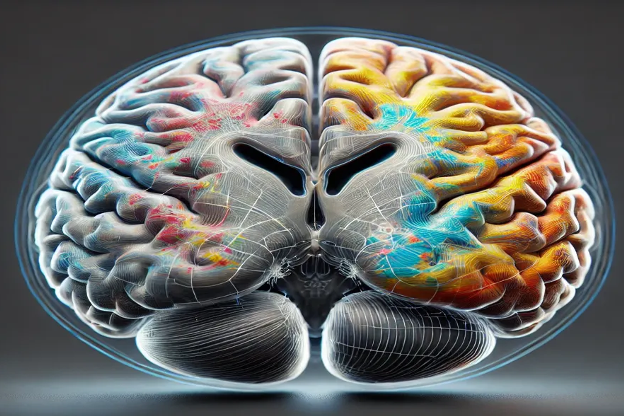
Neuroimaging, a fusion of neuroscience and medical imaging, serves as a window into the living brain, enabling us to observe both its structural complexity and its dynamic functional processes. Think of neuroimaging as a camera that not only captures static pictures of the brain’s anatomy but also films its activities, allowing us to understand how the brain operates as we think, learn, and interact with the world around us.
In the last 30 years, neuroimaging technologies such as Magnetic Resonance Imaging (MRI) and Functional MRI (fMRI) have revolutionised our understanding of brain function. These tools allow us to explore how various brain regions communicate and collaborate to support cognitive processes, memory formation, and motor skills. Neuroimaging doesn’t just map the brain’s physical structure; it enables the study of brain activity in real-time, providing critical insights into neurological health, cognitive functions, and disease progression.
Real-World Example: Learning and Brain Activity
Consider a beginner learning to play the piano. Early neuroimaging scans might show heightened activity in the motor cortex, as the person struggles with the mechanics of finger movement and hand-eye coordination. Over time, as muscle memory develops, activity in the motor cortex decreases, while regions involved in memory retrieval and coordination become more active. This shift in brain activity reflects the learning process and neural plasticity — the brain’s ability to adapt to new tasks.
The Importance of Neuroimaging in Modern Science
Before the advent of neuroimaging, studying the brain relied on post-mortem examinations or observing patients with brain injuries, which provided limited insights into healthy brain function. Today, neuroimaging has opened new avenues for studying living brains in real-time. Researchers can now observe how neural circuits support complex cognitive functions, how the brain changes over time, and how diseases impact brain structures. This has transformed not only neuroscience but also psychology, medicine, and even artificial intelligence.
Key Impacts of Neuroimaging:
- Medical Diagnosis: Neuroimaging is indispensable in diagnosing neurological diseases such as Alzheimer’s, Parkinson’s, and multiple sclerosis. Brain scans can reveal tissue degeneration in Alzheimer’s, identify abnormal dopamine-producing regions in Parkinson’s, or highlight white matter lesions in multiple sclerosis. Such data allows for earlier diagnoses and more effective treatments.
- Research on Brain Plasticity: Neuroimaging enables the study of brain plasticity — the brain’s ability to reorganize itself by forming new neural connections. For example, researchers can use fMRI to observe how the brain compensates for injuries by recruiting other regions to take over lost functions, offering insight into rehabilitation strategies.
- Intervention and Treatment: Doctors use neuroimaging to guide interventions. In epilepsy treatment, for instance, neuroimaging helps pinpoint the brain region causing seizures, which allows for targeted surgery or localized treatments. Similarly, deep brain stimulation (DBS) for treating Parkinson’s disease uses imaging to map out the areas that will be stimulated to improve motor function.
Expanded Discussion on Treatment Development
Beyond diagnosis, neuroimaging plays a transformative role in the development of new therapies. For conditions like Alzheimer’s disease, neuroimaging helps monitor the effects of drugs designed to slow down or halt neurodegeneration. By tracking changes in brain volume or measuring cortical thickness over time, researchers can assess the effectiveness of a treatment. This iterative process allows for faster refinement of therapeutic strategies.
How Does Neuroimaging Work?
Neuroimaging involves advanced equipment and algorithms to generate detailed images of the brain. Each technique produces specific types of images, providing insights into different aspects of brain structure and function. Let’s explore the most commonly used neuroimaging techniques and their applications:

1. Magnetic Resonance Imaging (MRI)
MRI produces high-resolution images of the brain’s structure by using powerful magnets to manipulate the magnetic fields around water molecules in the brain. The resulting images allow researchers to study different brain regions’ sizes, shapes, and structural integrity.
Expanded Example: Cortical Thickness and Its Significance
Cortical thickness refers to the distance between the white matter and the brain’s surface (cortex). This measure is vital for studying brain development and aging. For instance, increased cortical thickness in the prefrontal cortex is associated with enhanced executive function, attention, and decision-making. Conversely, thinning of the cortex in specific areas is linked to neurodegenerative diseases like Alzheimer’s. By measuring cortical thickness over time, scientists can track both normal brain development and the early onset of diseases.
2. Functional Magnetic Resonance Imaging (fMRI)
fMRI provides a dynamic view of brain activity by measuring changes in blood flow. When a region of the brain becomes active, it requires more oxygen, which leads to increased blood flow to that area. fMRI can track these changes, enabling researchers to map brain activity in real time.
During language processing tasks, fMRI reveals increased activity in areas like Broca’s area (involved in speech production) and Wernicke’s area (critical for language comprehension). By mapping these activations, scientists can study how different parts of the brain work together to enable complex cognitive tasks such as reading, speaking, and understanding language. fMRI has also been used to study language acquisition in children and how the brain adapts to learning new languages.
3. Diffusion Tensor Imaging (DTI)
DTI is a specialized form of MRI that focuses on the brain’s white matter — the tracts or “highways” that connect different brain regions. By tracking the diffusion of water molecules along these tracts, DTI provides insights into the integrity and connectivity of brain pathways.
Expanded Insight: Understanding Brain Connectivity
DTI is instrumental in understanding how brain diseases affect connectivity. In conditions like multiple sclerosis, DTI can show how the disease damages the white matter tracts, disrupting communication between different brain regions. Such visualizations help clinicians assess the extent of damage and track disease progression. DTI also plays a key role in connectomics, a growing field focused on mapping all the connections in the brain to understand how these pathways support cognitive functions.

Key Files Generated from Neuroimaging
When neuroimaging data is collected, it generates a variety of files, each offering unique information about the brain. These files are essential for different stages of analysis and help researchers measure brain structure, function, and connectivity.
1. Raw MRI Files (.mgz or .nii)
Raw MRI files store unprocessed data directly from the MRI scanner, providing a 3D image of the brain. These files are the foundation for all subsequent analyses and contain detailed information about the brain’s structure before any processing.
2. Skull-Stripped Brain File
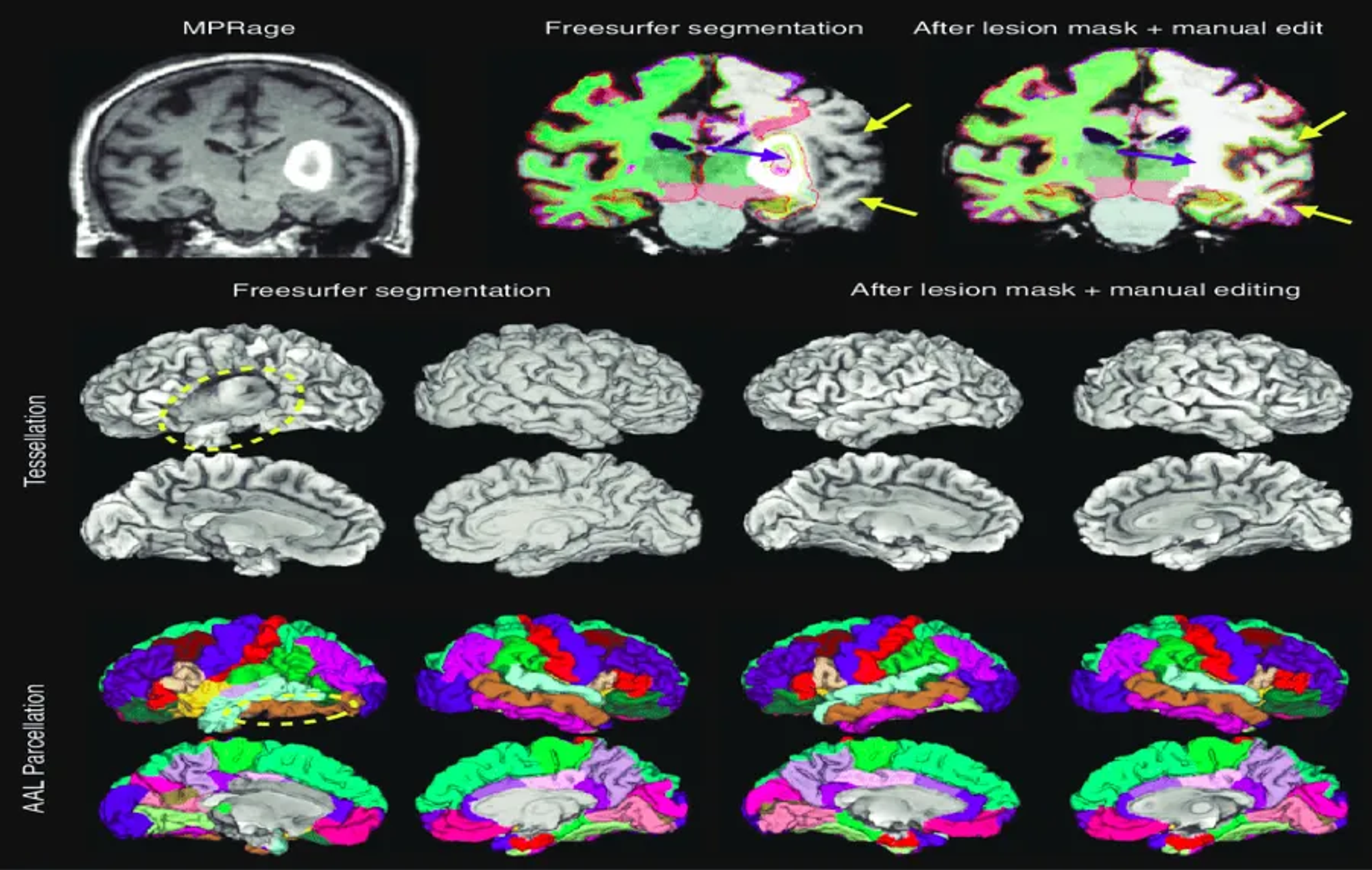
Once raw data is collected, the skull and other non-brain tissues are removed, resulting in a brain-only image. This skull-stripped file improves the accuracy of measurements like brain volume and cortical thickness by eliminating non-relevant data.
3. Cortical Surface Reconstruction Files
These files capture a 3D model of the brain’s surface, including its intricate folds (gyri and sulci). Researchers use these models to measure the surface area and depth of the folds, which have been linked to cognitive abilities.
Example: Cortical Surface Analysis
Studies show that individuals with higher cognitive abilities tend to have a more folded cortex, particularly in areas related to problem-solving and memory. Neuroimaging techniques like cortical surface reconstruction allow scientists to quantify these folds and study their relationship with cognitive function.
4. Diffusion Tensor Imaging (DTI) Files
DTI files store data about the brain’s white matter tracts. These files provide detailed maps of the brain’s internal connections and help researchers study how different brain regions communicate.
Example: Studying Connectivity in Autism
In people with autism spectrum disorder (ASD), DTI has shown alterations in white matter tracts, suggesting that disrupted connectivity between brain regions could underlie some of the cognitive and social challenges associated with the condition.
The Role of Computational Neuroscience in Interpreting Neuroimaging Data
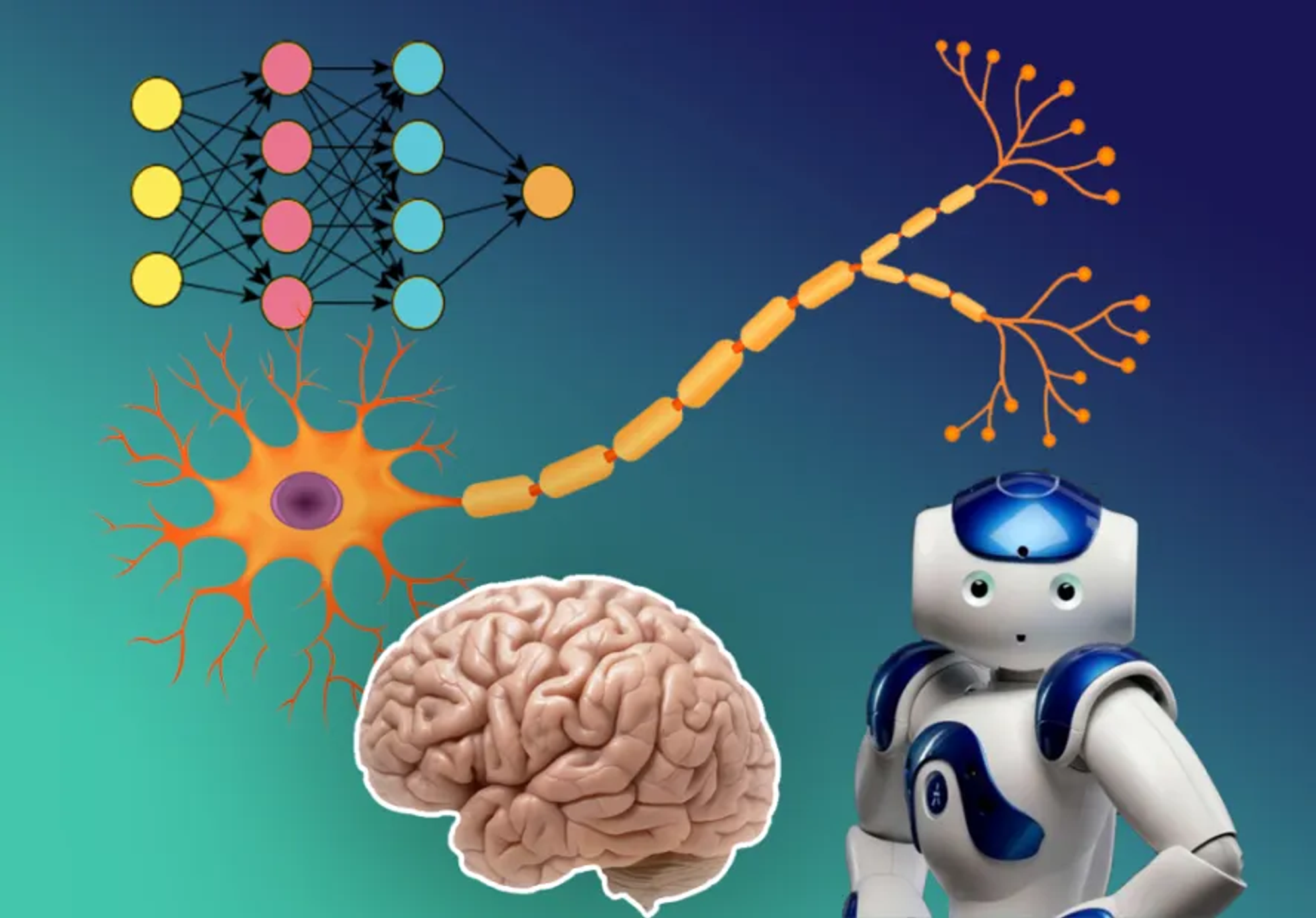
Neuroimaging data is vast and complex, requiring sophisticated computational tools to make sense of it. Computational neuroscience uses mathematical models, algorithms, and simulations to interpret this data, helping us understand how the brain processes information and performs tasks.
1. Neural Networks and Brain Function
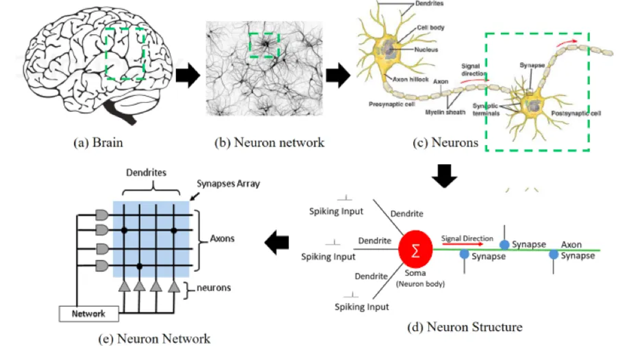
In computational neuroscience, researchers create models of neural networks that simulate how groups of neurons in the brain communicate and work together. These models can mimic brain functions like vision, memory, or motor control, allowing researchers to test hypotheses about how the brain operates.

Real-World Application: Visual Processing
Scientists use neural network models to simulate the brain’s visual system, from basic edge detection to complex object recognition. By comparing these simulations to neuroimaging data, researchers can test how accurately their models reflect real brain processes, offering insights into how the brain interprets visual stimuli.
2. Machine Learning and Neuroimaging
Machine learning algorithms are increasingly used to analyze neuroimaging data. These algorithms can detect patterns in the data and make predictions about brain function or health status. For example, machine learning models trained on fMRI data can predict which brain regions will activate during specific tasks, helping scientists understand the relationship between brain structure and function.
Expanded Example: AI-Assisted Diagnosis
AI models trained on neuroimaging data can help diagnose neurodegenerative diseases like Alzheimer’s by identifying subtle patterns in brain scans that are invisible to the human eye. In recent studies, AI models have been able to predict Alzheimer’s onset years before clinical symptoms appear by analyzing changes in brain volume and connectivity.
Challenges and Ethical Considerations
As with any technology, combining neuroimaging with AI presents both opportunities and challenges.
1. Data Complexity
Neuroimaging generates enormous amounts of data, and understanding this data requires advanced computational techniques. Small variations in data collection can significantly affect results, making it challenging to analyze brain scans consistently across different studies.
2. Ethical Concerns
The collection and use of brain data raise serious ethical concerns. Brain scans are deeply personal, and questions about data privacy and ownership remain unresolved. For example, should insurance companies have access to brain data to assess neurological health risks? Current regulations like GDPR and HIPAA aim to protect personal health data, but more robust safeguards may be necessary as neuroimaging becomes more prevalent.
3. Computational Power
Both neuroimaging and AI require significant computational resources. Processing real-time brain data — especially from techniques like EEG or fMRI — requires enormous processing power. Advances in quantum computing and cloud-based AI may eventually alleviate these challenges, but for now, computational capacity remains a major hurdle.
Conclusion
Neuroimaging, paired with computational neuroscience and AI, holds immense potential for deepening our understanding of the human brain. From improving medical diagnoses to unraveling the mysteries of cognition, these technologies offer unparalleled insights into brain function and health. However, the field faces challenges in managing data complexity, ensuring ethical use, and providing sufficient computational resources. As technology advances, we may see a future where personalized AI systems, informed by neuroimaging, adapt seamlessly to our unique cognitive styles, enhancing everything from healthcare to education.
